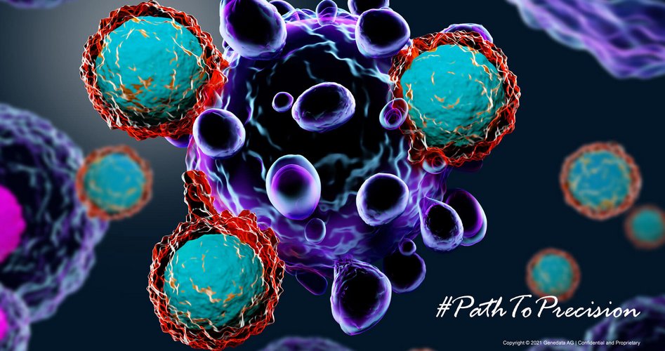
Path to Successful Cell Therapy: Going Beyond a Biomarker-Driven Strategy
November 30, 2021
Justyna Lisowska
Despite rapid and intensive developments of new cell therapy modalities, cell immunotherapies still face several challenges that limit their wider use in the clinic. Up to date, a small number and class of malignancies have been successfully treated with this approach, the patient response rate is highly variable1, and a considerable number of patients still relapse by month 12 following treatment administration2. These therapies also expose patients to severe adverse events due to heightened immune reactions.
As objective quantifiable biological indicators of pathophysiological and physiological processes, biomarkers can be used to predict and monitor the efficacy of a given treatment and the incidence of adverse events. They can also facilitate the development of novel therapeutic agents and improve existing treatments by determining the best therapy responders. The identification of robust cell therapy biomarkers could enable better management of immune-mediated toxicity, prediction of efficacy or relapse, ultimately accelerating clinical development and improving healthcare outcomes.
However, contrary to other immune-oncology treatments, the desired therapeutic effect of cell immunotherapies depends not only on patient-specific molecular features but also on the inherent attributes of the cellular product and its interaction with the host environment. Therefore, to ensure safe and efficacious cell therapy, we need to go beyond a biomarker-driven strategy. We need to understand the whole multi-dimensional system by unravelling interconnections between the functional and phenotypic properties of these living drugs, disease characteristics (including tumor & tumor microenvironment profile), patient immune systems as well as their genetic, epigenetic, metabolic, and other molecular features.
Management of Immune-Mediated Toxicity
Cytokine Release Syndrome (CRS) or the “cytokine storm” is the most common side effect following cell therapy treatment limiting durable cell therapy-mediated disease remission. The cytokine storm is a systemic inflammatory reaction triggered by inflammatory cells (e.g., T cells, and macrophages) and the immune mediators released upon their activation in response to the receptor’s binding to specific antigens. Therefore, the expression level of receptor engagement surface signatures (e.g.CD3 and CD8) could be indicative of immune-mediated toxicity.
While it is difficult to predict the incidence of CRS ahead of time, we can monitor the reactivity of a patient’s immune system during treatment to alleviate the consequences of such an adverse event. Evaluating the systemic level of biological factors involved in this immune reaction such as T cell activating cytokines (IL-6/ INF-gamma, SIL-2R alpha, sIL-6R, and GM-CSF), macrophage activating cytokines (IL-6, IL-1Ra, IL-10, IP-10, and sIL-6R), or macrophage attracting chemokines (MCP, IP-10, and IL-8) using multiplex bead array technology provides a useful means of early identification of such toxicity, allowing its better management.1,3
A more severe form of CRS is often accompanied by coagulopathy, tumor lysis syndrome (TLS), hemophagocytic lymph-histiocytosis/macrophage-activation syndrome (HLH/ MAS), or organ dysfunction. Therefore, other serum biochemical parameters involved in these disorders such as angiopoietin-2 (Ang2), von Willebrand factor (vwF), D-dimers, C-reactive protein (CRP), ferritin, LDH, aminotransferase (AST), alanine aminotransferase (ALT), blood urea nitrogen (BUN), and creatinine could be used as potential indicators of cell therapy-induced toxicity. Systems biology-based platforms enabling molecular array- and proteomics-based analyses, as well as high throughput multiplex-bead array-based assays would be highly beneficial to monitor modulations in the level of multiple immune factors following cell therapy administration.3
Ensuring Long-Term Cell Therapy Efficacy
Patient Profiling & Surveillance
Another concern regarding cell immunotherapies is their variable long-term efficacy. Patient-specific features associated with a primary disease or previous treatments can considerably impact the response to cell therapy. For instance, while higher tumor mutational burden, indicative of tumor immunogenicity, increases the likelihood of positive therapy response, patient exposure to checkpoint inhibitors (e.g., anti-CTLA-4 treatment) before receiving cell therapies limits long-term therapy benefit1.
The presence of specific tumor antigens is necessary for immune cells to successfully recognize and eliminate cancerous cells and thus, prerequisite for effective cell therapy. A minimum threshold of antigen expression is required to ensure long-term CAR-T cell functionality4, while impaired interaction between the antigen receptor and its target (due to loss or dysfunction of either one), leads to disease relapse5. Continuous monitoring of tumor antigen expression is thus of high importance to ensure long-term therapeutic benefit. Determining genetic alterations underlying disrupted antigen expression and their identification with tools like next-generation sequencing (NGS) could inform about the likelihood of disease recurrence ahead of time.
In addition to post-treatment antigen loss, the presence of antigen-negative clones within the tumor may be responsible for the immune escape-mediated relapse. The identification of such cell populations, displaying genetic alterations, or alternatively spliced T-cell targets within the tumor would be helpful to mitigate the risk of relapse. Post-treatment monitoring of minimal residual disease (MRD) with flow cytometry, polymerase chain reaction (PCR), or NGS - based methods may be used to inform on the presence of cell subpopulations resistant to cell therapy. Finally, cell therapy efficacy may be significantly limited by tumor-mediated strategies to bypass the anti-tumor activity of the immune system. Hence, gaining insight into immunosuppressive mechanisms developed within the patient’s tumor microenvironment may be useful to assess the potency of cell therapy. Besides verifying checkpoint molecule expression on cancerous cells, checking the presence and abundance of myeloid-derived suppressor cells within the tumor might be beneficial. Assessing the level of immune mediators capable of mitigating tumor-based immunosuppression such as INF – γ, MIP-1, IL-8, granzyme B, IL-17, and IL-5, may also help to evaluate the potential outcome of cell therapy.6
Besides profiling a patient’s tumor, an improved understanding of a patient’s immune system would be of high importance to predict and assess the potential therapeutic outcome as well as mitigate the risk of adverse events. This includes the identification of pre-treatment immune fitness markers (baseline T cell level, proliferation, differentiation, and survival potential) as well as monitoring immune reactions following infusion (cytokine production, tumor lysis) and its systemic impact.
Determining Cell-Specific Attributes
Cell therapy efficacy depends on the characteristics of the biological material used. The phenotype, functional competence of cells before engineering, as well as their in vitro and in vivo features post-infusion (e.g., expansion, persistence, and bioactivity in response to tumor antigens) all determine the ability of the cell therapy to fulfill its function. The exact cell phenotypes and mechanism(s) associated with therapeutic benefit are difficult to determine given the heterogeneity of cell therapy products. Manufacturing processes involving stimulation with cytokines or growth factors, genetic engineering, or in vitro amplification, vary in terms of reagents, protocols, or laboratory conditions used. These factors contribute significantly to the outcome of the product. Moreover, the starting material considerably differs from donor to donor due to individual genetic and epigenetic backgrounds, physiological conditions, underlying comorbidities, or treatment history. Having all these heterogeneous data under one roof integrated and harmonized would allow to decipher key attributes of optimal donors, facilitating cell product standardization.
In the pursuit of an optimized cell therapy product, phenotypic characterization of T cells before infusion based on surface molecule expression provides insight into their bioactivity and thus, potency as a drug. Several studies have shown that less differentiated cells have higher proliferation potential, better persist within the patient, and are more effective in fighting tumor cells6. Therefore, assessing markers of T cell differentiation (CD45 RA or RO, CD62L, CCR7, CD27, CD28) using multi-channel flow cytometry would help to determine a functional cell therapy product. Early memory CAR-T cell subsets are usually found in complete responders. Hence, functional, and genomic characteristics that dictate cell memory phenotype, such as IL-6, STAT3 signatures (IL-6/IL-17/IL-22/IL- 31 and CCL20), can also be indicative of therapy efficacy1. In contrast, T cells with upregulated genes of end effector differentiation, glycolysis, and apoptosis correlate with poor clinical outcomes1. The overexpression of PD1 checkpoint molecules on post-manufactured CAR-T cells is predictive of limited T cell expansion, cytokine release, and cytotoxicity, while a high level of LAG3 and TIM3 is associated with T cell exhaustion and overall poor cell therapy response1.
Monitoring Cellular Fitness
Selecting cells with optimal attributes for the treatment increases the probability of therapeutic success. Nevertheless, long-term therapy effectiveness can only be ensured if cellular fitness is maintained within the patient’s body. Patients may experience disease recurrence due to limited CAR-T cell persistence and expansion. To prevent this, it is advisable to monitor the presence and behavior of the cellular product on an ongoing basis. Flow-cytometry based measurement of cell surface markers, such as specific TCR α/β pairs, a variable domain of the TCRβ chain (Vβ) or CAR molecules can be used to determine the quality and quantity of effector cells post-implantation3. High-throughput and deep sequencing of TCR variable (V) and CDR3 domains, as well as the detection of transgene amplification by quantitative PCR of peripheral blood mononuclear cells (PBMC) samples, can also be used to monitor the presence and/or expansion of infused cells3.
Finally, the expression profile of activation markers (CD25, CD127, CD57, and CD137) or certain cytokines might be a useful indicator of the functional T cell product. Looking at IFN-γ, IL-2, IL-4, and IL-10 can help assess T cell activity following antigen engagement7. Also, it has been shown that patients treated with CD8+ CAR-T cells expressing a high level of TNFα, and a low level of TIM-3 experience longer remission6.
Empowering Scientific Insights with a Technological Solution
The recent FDA clinical hold of Allogene’s AlloCAR-T product following unexpected chromosomal abnormalities indicates the complexity behind cell immunotherapy8. Such examples highlight the importance of characterizing biological factors that lead to successful clinical products. It also demonstrates the need to understand and monitor the dynamic interplay between the living drug, tumor, immune system, and other patient-specific molecular and physiological characteristics over time during the clinical trial and beyond.
However, deciphering this entire complex system from a variety of data and identifying relevant biological indicators that could mitigate toxicity and efficacy concerns is extremely difficult as simultaneous real-time characterization at different molecular levels (genomic, transcriptomic, proteomic, and cytomic) is required. To grasp the full biological picture and derive actionable insights, the relationships between changes in such characteristics need to be identified. This requires data integration and systems-biology-based analyses which can only be done within a performant informatic platform capable of storing, processing, and analyzing ever-increasing amounts of new multimodal data on an ongoing basis. Such integrative analyses on high-throughput multi-omics data can be performed only if data standardization and harmonization steps are ensured. Once the datasets are integrated, advanced bioinformatics and computational tools can come into play to enable downstream analysis. The use of Artificial intelligence (AI), specifically machine learning, is useful in the generation of predictive models and the validation of biomarkers. As cell therapy development involves rapid cycles of innovation and the accumulation of new data types from diverse technologies, innovative solutions with a high level of flexibility are required. A data-agnostic, agile system capable of adapting to the changing needs of biopharmaceutical and biotechnological companies would accelerate the advancement of cell therapies.
Genedata Profiler® is a scalable enterprise software solution that addresses complexities involved in handling an increasing amount of diverse data through automated data processing, harmonization, and integration. By equipping scientists with an advanced performant analytical layer to run complex analytics in a validated environment, clinically relevant markers of toxicity and efficacy can be deciphered from the wealth of translational and clinical data. Moreover, enabling real-time data visualization through customized dashboards, developed using integrated 3rd party tools, accelerates and facilitates data-driven decision making. Finally, critical for innovation is the facilitation of efficient collaboration. In addition to custom visualization available on-demand, the Genedata Profiler platform provides self-service access to data streamlining knowledge sharing and fostering collaboration between cell therapy research and manufacturing teams, as well as external partners.
Learn More About Genedata Profiler
Similar Articles:
- Cell Therapy: The Potential for Effective Precision Medicine in Oncology and Beyond
- The Promises and Challenges of Autologous and Allogeneic Cell Therapies
- Leveraging Data Gathered on the Journey to Successful Cell Therapy
- Podcast: The Power of Data in Cell therapy Development
References:
- Han et al., Defining precision cellular immunotherapy—seeking biomarkers to predict and optimize outcomes of T cell therapies in cancer. Precision Cancer Medicine, 2019
- Du et al., Biomarkers in individualized management of chimeric antigen receptor T cell therapy. Biomarker Research, 2020
- Kalos M., Biomarkers in T cell therapy clinical trials. Journal of Translational Medicine, 2011
- Walker AJ, et al. Tumor antigen and receptor densities regulate efficacy of a chimeric antigen receptor targeting anaplastic lymphoma kinase. Mol. Ther., 2017.
- Shah NN, Fry TJ. Mechanisms of resistance to CAR T cell therapy. Nat Rev Clin Oncol., 2019
- Hong, R. et al., Biomarkers for Chimeric Antigen Receptor T Cell Therapy in Acute Lymphoblastic Leukemia: Prospects for Personalized Management and Prognostic Prediction. Front. Immunol., 2021
- Stroncek DF et al., Global Transcriptional Analysis for Biomarker Discovery and Validation in Cancer and Hematological Malignancy Biologic Therapies. Mol Diagn Ther., 2009
- Caroll J. Allogene hit with FDA clinical hold after a patient experiences 'abnormality' in CAR-T cells — shares hammered Endpoints News, Oct 2021